Leader:
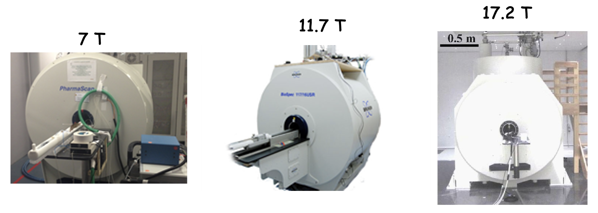
The “Small animal MRI systems” platform includes three active 7T, 11.7T and 17.2 MRI scanners provided by the Bruker BioSpin company, mainly dedicated to rodents and small animals imaging. The three scanners are equipped with anaesthesia systems calibrated for isoflurane that can be used both inside and outside the magnet. Respiration, temperature and electrocardiogram sensors continuously monitor the animal health, and the signals coming from these sensors can be used to synchronize the MRI acquisition. An air heater or a warm water circulation system ensure a stable body temperature, thanks to a closed loop algorithm based on the measured temperature of the animal. In addition, an isolated pulse stimulator, a programmable injector, an artificial ventilation system for rodents and a dissolved gas analysis machine are also available for specific experiments.
The 7T preclinical MRI scanner, running under the responsibility of S. Mériaux from the “MIDAS” research group, is a Pharmascan system purchased from Bruker BioSpin company. It is equipped with the AVANCE III electronics allowing imaging of proton/fluorine nuclei, as well as other X nuclei at lower Larmor frequencies. The proton/fluorine channel consists of 4 independent receive channels managed by the licensed GRAPPA parallel imaging package from Bruker. The high strength of the magnetic field gradients, capable of reaching 740 mT/m, allows performing diffusion MRI acquisitions at high b-values. The system is controlled by the Paravision software (version 6), including the standard imaging licenses together with the following additional imaging sequences: spectroscopy, diffusion tensor imaging, echo-planar imaging (EPI), parallel imaging, echo-navigated cardiac imaging, ultrashort TE and zero TE imaging. Moreover, the 7T scanner is equipped with a variety of radiofrequency coils: surface, phased-array and volume coils for rat and mouse brain imaging, X nuclei surface coils (13C, 19F, 31P) and a dual-resonance (1H/19F) volume coil. Finally, the 7T magnet is also equipped with a unique motorized system for focused ultrasound, dedicated to non-invasive transcranial sonications in rodent heads (co-developed with Image Guided Therapy company). This setup can be used for protocols of transient permeabilization of the blood-brain barrier, local tissue thermal ablation or ultrasound-induced neuromodulation.
The 11.7T preclinical MRI scanner, running under the responsibility of F. Boumezbeur from the “CICLOPS” research group, is the state-of-the-art system for biomedical imaging of rodents. In addition to its intense static magnetic field, this MRI system is equipped with high magnetic field gradients (760 mT/m) and a specific cryo-platform including a two-channel cryoprobe (cooled at 25K) dedicated for brain imaging of mice. The extreme cooling leads to a 250% enhancement in signal-to-noise ratio (SNR) compared to a similar radiofrequency (RF) coil operating at room temperature. Thus, optimal spatial and temporal resolution can be achieved. Moreover, this scanner is equipped with 4 reception channels for parallel imaging and a large array of dedicated RF coils for most preclinical applications: a set of surface coils, a 4-channel phased-array coil, volume coils for whole body rat and mouse imaging and dual-resonance (1H/13C, 1H/31P) coils for hetero-nuclear MR spectroscopy.
The 17.2T preclinical MRI scanner, running under the responsibility of L. Ciobanu from the “NeuroPhysics” research group, is one of the highest magnetic field horizontal bore preclinical system in the world. The clear bore inside the gradients (85 mm) allows the imaging of small animals (rats, mice). Besides the very high magnetic field, another feature of this system is the high strength of the magnetic field gradients, capable of reaching 1000 mT/m. With these gradients, it is possible to acquire very high spatial resolution images (25 µm isotropic). The 17.2T system is equipped with a variety of radiofrequency coils: surface, phased array and volume coils for rat and mouse brain imaging, as well as with microcoils with diameters between 700 µm and 2 mm for imaging small samples. X nuclei surface coils (13C, 31P) for spectroscopy studies are also available.
The system will be soon equipped with the AVANCE NEO electronics that offers improved shimming performances, faster gradient switching times and gradient linearity corrections ensuring better image quality. Secondly, a cryo-platform and a cryo-cooled deuterium coil (2H) will be purchased to open the way to novel explorations of metabolic events occurring in the brain. A helium recovery and liquefaction system will be installed to eliminate the need for helium supplies.