Leader:
The histology platform
The histology platform, running under the responsibility of F. Geffroy from the “MIDAS” research group, consists in a technical facility dedicated to the histopathological analysis of animal models currently under investigation in several preclinical projects. This facility provides all equipment and techniques required for histological data acquisition, starting from biological samples preparation until their analysis with fluorescence microscopy. To meet the users’ needs, the histology platform offers a wide range of experimental protocols:
-
organs harvesting, intracardiac perfusion
-
cryoprotection with sucrose, freezing with isopentane
-
cryomicrotomy to acquire histological slices
-
slices collection on glass slides before storage at -30°C or -80°C
-
set-up of immunohistochemical (simple or multiple) and histochemical stainings
-
visualization of histological stainings using bright-field or fluorescence microscopy. An AxioObserver Z1 microscope is available, with Zen 2 software) providing confocal-like images, together with an Imaris station to analyse images.
Histological studies performed after MRI studies are a classic way to confirm results obtained with MRI and to characterize the metabolic, functional and structural cellular organization of the organs (microstructure, active cells or dying cells, interactions between contrast agents and target, vasculature…) at a better resolution than MRI. The facility also offers the possibility to perform immunostaining on the tissue to map their microenvironment (inflammation, apoptosis, production of new cells, etc.), as well as to perform chemical staining to reveal iron core of nanoparticles. Clearing organs methods are commonly used to acquire data deeper in tissue.
The cell culture platform
The cell culture platform of NeuroSpin, running under the responsibility of F. Geffroy from the “MIDAS” research group, consists in a technical facility dedicated to the preparation of specific cell lines that are required to induce brain tumors in animal models currently under investigation in several preclinical projects. This facility provides all equipment and techniques required for cell culture:
-
a specific cryopreservation system for storing different cell lines of brain tumors (U87, C6, 9L, Gl261…)
-
a specific biological safety cabinet to handle cells in a clean working environment
-
a specific incubator to ensure cells growth in a strictly controlled CO2 environment
-
a specific binocular microscope for visualization of living cells
The laboratory also offers training courses for students and researchers to teach them how to use the laboratory’s various instruments, and how to carry out their projects in a secure environment that complies with current regulations.
Publications
The two most relevant ones (<5 years):
-
Jelescu, I.O., de Skowronski, A., Geffroy, F., Palomboe, M., Novikov, D. S., (2022) Neurite Exchange Imaging (NEXI): A minimal model of diffusion in gray matter with inter-compartment water exchange Neuroimage 119277 2022 https://doi.org/10.1016/j.neuroimage.2022.119277
-
Conti, A., Geffroy, F., Kamimura, H. A. S., Novell, A., Tournier, N., Mériaux, S. and Larrat, B. (2023) Regulation of P-glycoprotein and Breast Cancer Resistance Protein Expression Induced by Focused Ultrasound-Mediated Blood-Brain Barrier Disruption: A Pilot Study. International Journal of Molecular Sciences. 23, 15488 2022/ Int. J. Mol. Sci. 2022, 23, 15488. https://doi.org/10.3390/ijms232415488
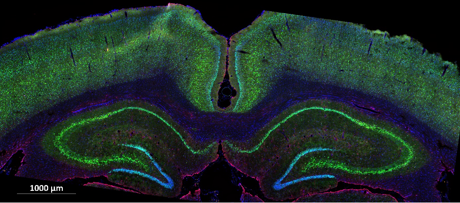
* The microphotography at the top of the page is a rat’s hippocampus, x20, Neurons revealed by NeuN immunostaining (green), astrocytes revealed by GFAP immunostaining (red) , nuclei revealed by DAPI staining (blue).
 Mouse cortex, x20 (MIP image, ROI), family of calcium binding proteins neurons among all others neuronal type, Neurons revealed by NeuN immunostaining (orange), interneurons expressing calcium binding protein revealed by immunostaining (Parvalbumin (red), Calbindin (blue) and Calretinin (green)).
 Alzheimer Rat’s cortex, x20, Amyloids plaques revealed by Thioflavine coloration (green), astrocytes revealed by GFAP immunostaining (red) , microglial cells revealed by IBA1 immunostaining (orange).
 Brain‘s mouse bearing human tumor (U87MG cells), x20, U87MG cells revealed by anb3 integrin immunostaining (orange), vessels revealed by CD31 immunostaining (green), microglial cells revealed by IBA1 immunostaining (red) and theranostic nanoparticles (magnetosomes) revealed by AMB1 immunostaining (blue).